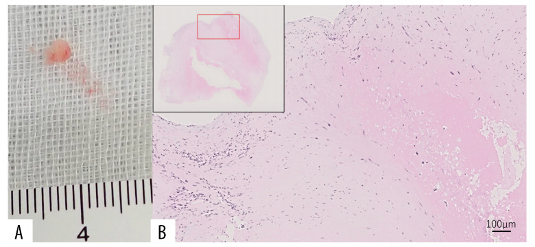
Figure 3.
(A) Macroscopy of the mitral vegetation in the tissue sample of the patient. (B) Hematoxylin & eosin stain of the histopathological mitral vegetation tissue showing an organized fibrin thrombus with no neutrophilic infiltration (×20 magnification), consistent with Libman-Sacks endocarditis.

