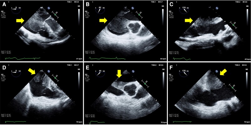
Figure 4.
Preoperative transoesophageal echocardiography showed an intra-cardiac mass (yellow arrows) in the right atrium and a thickened interatrial septum (A and B). The mass made contact with the broad area of the tricuspid valve, which was especially the posterior leaflet, during the diastolic phase (C). Postoperative transoesophageal echocardiography showed a reduction of mass, but the tumour involving the interatrial septum remained (D–F). AV, aortic valve.

