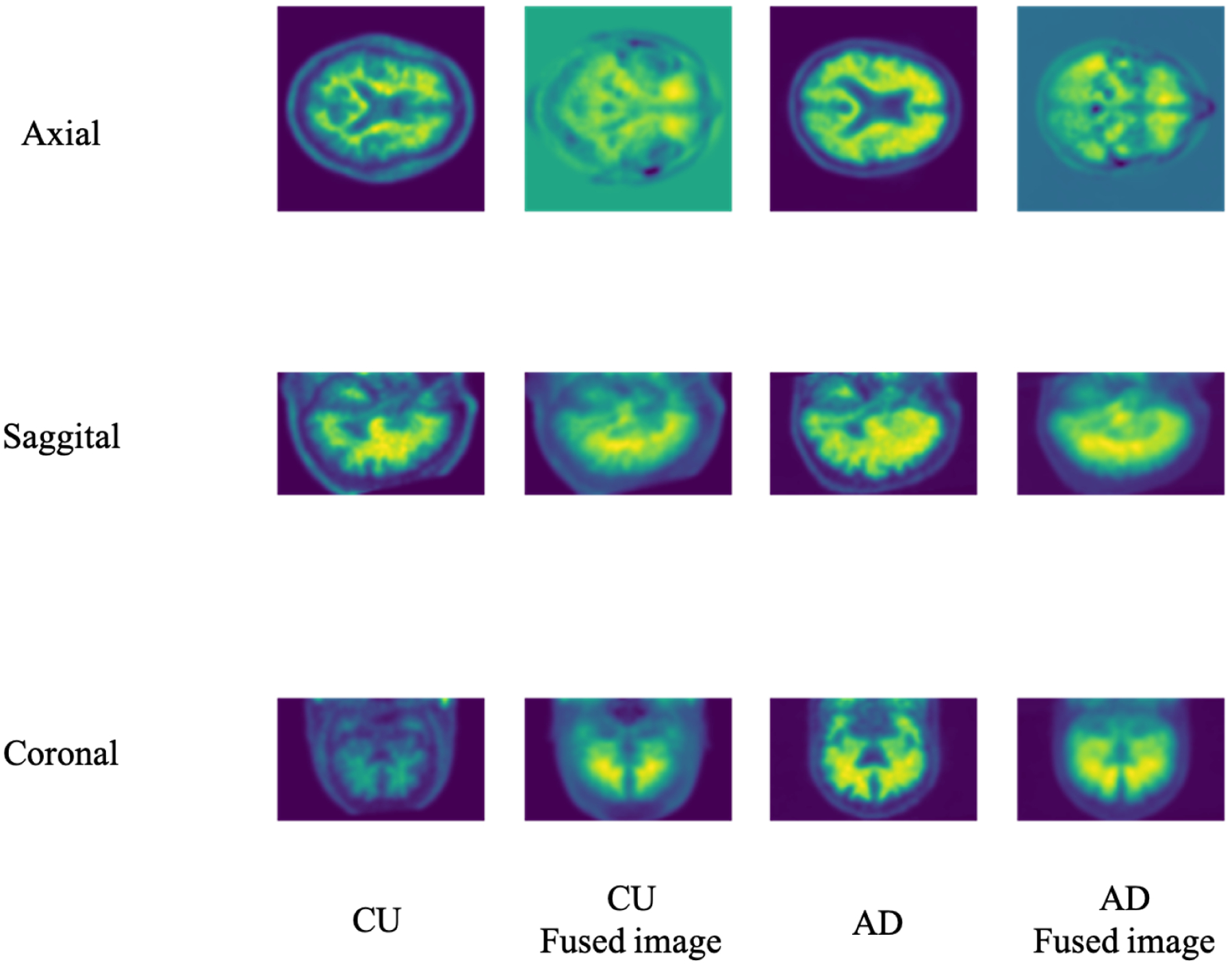
Figure 4.

The different views of the 3D Brain image slice and the corresponding 3D-to-2D fused image. Based on the axial, sagittal, and coronal views, the 3D PET image size is 96 × 160 × 160. The corresponding fusion images of PET under different views are different: 160 × 160 of axial view, 96 × 160 of sagittal view, and 96 × 160 of coronal view.
