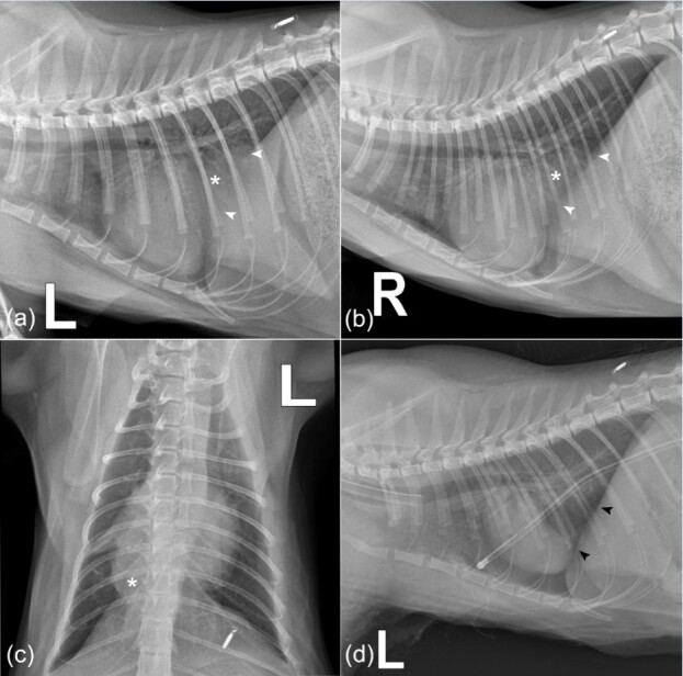
Figure 2. Thoracic radiographs of an 8-month-old neutered female domestic mixed breed cat with persistent coughing, mild asphyxia, nasal discharge, and physical activity intolerance. (a) Left lateral, (b) right lateral recumbency and (c) dorsoventral projections. Panel a-c shows an enlarged cardiac silhouette with cardiomegaly before surgical intervention. The cardiac silhouette merges with the diaphragmatic outline and loss of diaphragmatic border. There is a lobulated soft tissue opacity in the caudal thorax just right of the midline at mid-height, with a broad base to the diaphragm (asterisk). White arrowheads indicate the loss of diaphragmatic margins. The caudal vena cava is not visible. Left lateral (d) recumbency thoracic radiograph after herniorrhaphy and thoracic tube placement. Black arrowheads indicate the integrity of the diaphragm after the surgical procedure.

