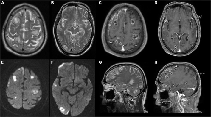
FIGURE 3.
Magnetic resonance image (MRI) of brain shows abscesses in bilateral frontal lobe, right temporo-parieto-occipital lobe, and right cerebellar hemisphere with ring contrast enhancement and perilesional edema. (A,B,G) T2-weighted imaging. (C,D,H) T2 contrast-enhancement imaging. (E,F) Diffusion-weighted MRI.

