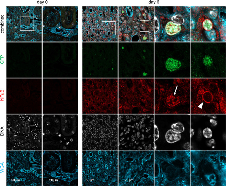
Fig. 4.
NFκB response in renal γδ T cells following S. aureus infection. TCRdH2BeGFP mice were infected with 107 CFU of S. aureus or remained without infection. Renal sections from mice at days 0 and 6 p.i. were stained with anti-GFP Ab to identify GFP+ γδ T cells (green; due to the histone 2B eGFP fusion protein, the staining is localized in the nucleus), anti-NFκB Ab (red), DNA (Hoechst, white), and wheat germ agglutinin (WGA, blue). Representative staining for sections from days 0 and 6 are shown (original magnification ×600). Large magnifications on the right show nuclear GFP+ γδ T cells with nuclear (arrow) and perinuclear (arrow head) NFκB staining. Additional sections for days 3 and 14 are presented in SI Appendix, Fig. S5. Sections are representative for five to seven mice per time point.

