Fig. 5.
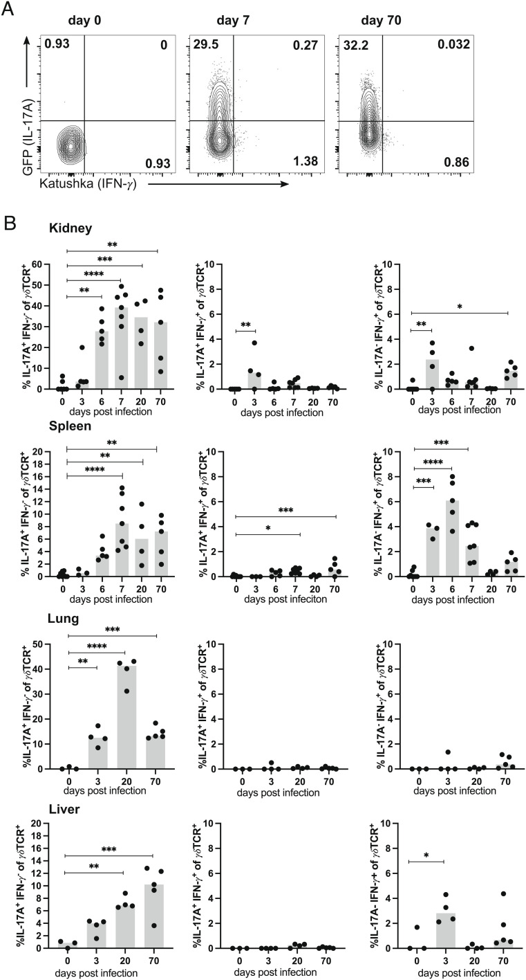
Renal γδ T cells retain IL-17A production during chronic S. aureus infection. Foxp3RFP×Il17aeGFP×IfngKat mice were infected with 107 CFU of S. aureus or remained without infection. At the indicated days p.i., cells were isolated from the spleen, kidney, lung, and liver, and CD45iv− γδTCR+ CD3+ cells were directly analyzed for cytokine reporter proteins. Three minutes prior to collecting of organs, mice received i.v. fluorochrome-conjugated anti-CD45 mAb to label vascular cells. (A) Representative dot plots for Katushka (IFN-γ) and GFP (IL-17A) expression in renal γδ T cells. (B) Percentages of IL-17A+IFN-γ−, IL-17A+IFN-γ+, and IL-17A−IFN-γ+ γδ T cells in organs. Representative result of two independent experiments with four to seven mice per group and time point. Symbols represent individual mice, and bars show median values. Statistical analysis was performed by one-way ANOVA test and Dunnett’s multiple comparisons posttest.

