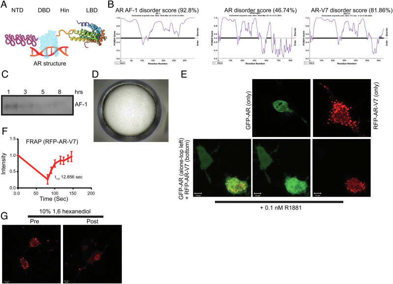
Fig. 1.
AR AF-1 and AR-V7 exhibit disordered protein characteristics. (A) AR structure (created using biorender.com; for illustration purpose only). (B) Pondr score. Pondr (www.pondr.com) disorder score for AR AF-1, AR full-length, and AR-V7. (C) Time-course for stability of recombinant-purified AR AF-1 (141 to 486). Purified recombinant AR AF-1 protein (5 ng) was incubated for the indicated time points at room temperature. Proteins were fractionated on SDS-PAGE and AR western blot was performed with an antibody that binds to the AF-1 region (AR-441, against AR fragment 302 to 318). (D) Recombinant-purified AF-1 (141 to 486) forms LLPS. Purified recombinant AR AF-1 (1 mg/ml) was seeded in a buffer (1:1 silver bullet E4 (1% w/v protamine sulfate, 0.02M HEPES pH 6.8) (HR2-096) + 30% PEG-3350 + 0.05 M HEPES pH 6.8) at 14 °C for 6 h and imaged using a phase contrast microscope. (E) AR-V7 forms molecular condensate in cells. COS7 cells were transfected with 1 µg turbo-red AR-V7 or GFP-AR or both. Twenty-four hours after transfection, the cells in DME + 5%csFBS without phenol red were treated with 0.1 nM R1881. Cells were imaged using a fluorescent confocal microscope 24 h after treatment. (F) FRAP. COS7 cells were transfected with 0.25 µg RFP-AR-V7. Twenty-four hours after transfection, the cells were fed with DME + 10% FBS. Forty-eight hours after transfection, selected regions were photobleached, and the recovery was monitored. Intensity of selected regions is provided as line graph (half-life (t1/2) is derived from an average of four independent measurements; represented as average ± SE). (G) 1,6 hexanediol dissolves the condensates. COS7 cells were transfected with RFP-AR-V7. Live cells in DME + 10% FBS were imaged before and 10 min after 10% 1,6 hexanediol addition. FRAP-fluorescence recovery after photobleaching; LLPS, liquid–liquid phase separation; NTD, N-terminal domain; DBD, DNA-binding domain; LBD, ligand-binding domain; RFP, red fluorescent protein; GFP, green fluorescent protein.

