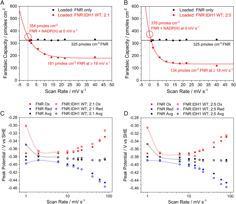
Fig. 4.
Scan rate-dependent Faradaic capacity (A and B) and trumpet plots (C and D) from thin-film voltammetry experiments using electrodes coloaded with FNR and wild-type IDH1 compared to an FNR-only electrode. At low scan rates, the NADP(H) carried in with IDH1 can be detected; peaks collapse to the FNR-only signal at high scan rates. (A and B) Scan rate-dependent coverage plots fitted with an asymptotic exponential equation to allow extrapolation to 0 mV/s. (C and D) Trumpet plots showing the changes in oxidation and reduction peak potentials as a function of scan rate (see trend lines). Conditions: stationary (FNR + IDH1)@ITO/PGE electrode (except for FNR-only data, which did not contain IDH1), electrode area 0.06 cm2, temperature 25 °C, volume 4 mL, pH = 8 (100 mM HEPES), and enzyme loading ratios (molar): (A and C): FNR/IDH1; 2/1; (B and D): FNR/IDH1; 2/5.

