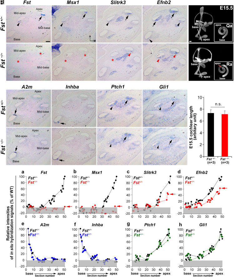
Fig. 1.
Specification of apical cochlear regional identity is compromised in Fst−/− cochleae. (A–P) In situ hybridization analysis with apical (Fst, Msx1, Slitrk3, and Efnb2) and basal (A2m and Inhba) regional markers and readouts of SHH signaling (Ptch1 and Gli1) in Fst+/− and Fst−/− embryos at E15.5. Arrows and arrowheads indicate relatively strong and weak expression, respectively. Red asterisks and red arrowheads indicate absence or down-regulation of expression, respectively. (Q–S) Paint-fill analysis of the Fst−/−inner ear (Q, R) and cochlear lengths of control and Fst−/−at E15.5(S). (T) Relative in situ hybridization signal intensities along the cochlear duct in Fst+/− and Fst−/− embryos. Red arrows indicate the apical end of the cochleae of Fst−/− embryos. Gray boxes below 0% in each graph indicate background signal. Representative graphs are presented from one Fst+/− and one Fst−/− cochlea for each gene. Additional samples are in SI Appendix, Fig. S1. The scale bar in A, 100 μm, also applies to B–P. The scale bar in Q, 100 μm, also applies to R.

