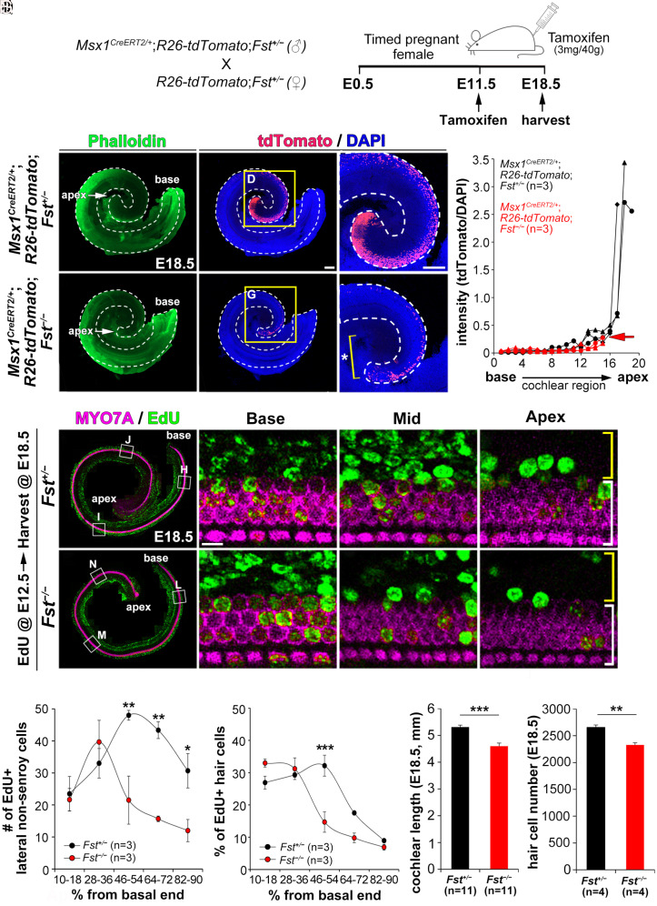
Fig. 2.
Failure of apical cochlear expansion caused by reduced cell proliferation in Fst−/− cochleae. (A–H) Fate-mapping of Msx1-lineage cells using Msx1CreERT2/+; R26-tdtomato mice in the presence (Fst+/−) or absence (Fst−/−) of FST. After tamoxifen was injected into pregnant female mice at E11.5, the embryos were harvested at E18.5 (A). Quantification of tdTomato-fluorescence intensity along the cochlear duct (H). (I–R) Comparing EdU-labeled cells in Fst+/− and Fst−/− embryos. After EdU was injected into pregnant female mice at E12.5, EdU-labeled cells (green) were counted among the lateral non-sensory cells (yellow brackets) and MYO7A-positive hair cells (white brackets) (I–P). The number of EdU-positive cells in the lateral compartment (Q) and the percentage of EdU-positive hair cells (R) were plotted against the distance along the cochlear duct divided into five regions from base to apex. (S–T) Comparisons of cochlear length and hair cell number between E18.5 Fst+/− and Fst−/− embryos. Data in Q–T are presented as means ± SE. Statistical comparisons were determined via two-way ANOVA for the EdU analysis and unpaired t tests with Bonferroni corrections for the cochlear length measurements (*P < 0.05, **P < 0.01, and ***P < 0.001). The scale bar in C, 200 μm, also applies to B–C, E–F, and I–M. The scale bar in D, 200 μm, also applies to G. The scale bar in J, 10 μm, also applies to K–L and N–P.

