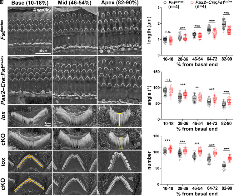
Fig. 3.
Loss of apical region stereocilia morphology in 4-wk-old Fst cKO mutants. (A–R) Scanning electron micrographs of the organ of Corti from control (lox) (Fstlox/lox) and inner ear-specific Fst cKO (Pax2-Cre; Fstlox/lox) mice. The lengths of the outer hair cell stereocilia were measured at the vertex of the V-shaped hair bundles from the lateral side (G–L). The angle of the V-shaped stereocilia and the number of stereocilia per outer hair cell were measured using a top-down view (M–R). The scale bar in A, 10 μm, also applies to B–F. The scale bar in G, 1 μm, also applies to H–R. (S–U) Quantification of the length, angle, and number of outer hair cell stereocilia along the tonotopic axis in each of the five cochlear regions beginning with the basal end. These represent the base (10 to 18%), mid-base (28 to 36%), mid (46 to 54%), mid-apex (64 to 72%), and apex (82 to 90%) regions. Statistical comparisons were determined via two-way ANOVA with Bonferroni corrections for multiple comparisons (n.s., non-significant, **P < 0.01, and ***P < 0.001).

