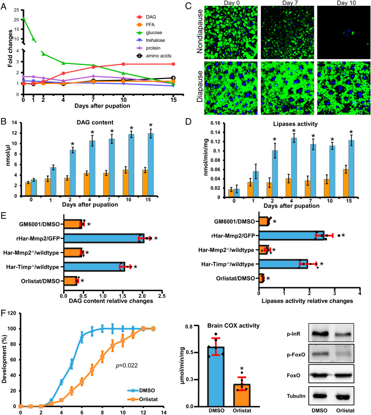
Fig. 4.
Mmp-induced fat body cell dissociation induces lipid mobilization. (A) Relative changes in diglyceride, free fatty acids, glucose, trehalose, total protein, and total amino acids in hemolymph from day 0 to 15 d after pupation in nondiapause pupae. (B) Developmental pattern of hemolymph diglyceride content in nondiapause and diapause pupae. (C) BODIPY staining of lipid droplets (green) in the fat body of nondiapause pupae. The nuclei were stained with DAPI (blue). (D) Developmental patterns of fat body lipases activity in nondiapause and diapause pupae. (E) Relative changes in hemolymph diglyceride content and fat body lipases activity in GM6001-injected pupae (ND-D7), pupae injected with recombinant Har-Mmp2 (NP-D1), Har-Mmp2−/− mutant pupae (ND-D7), Har-Timp−/− mutant pupae (ND-D1), and the lipases inhibitor Orlistat-injected pupae (ND-D7). (F) Delayed pupal development, decreased brain COX activity, and decreased levels of phosphorylated InR and FoxO in nondiapause pupae caused by Orlistat injection. Bars represent the mean ± SEM. Significant differences were calculated using Student’s t test (*P < 0.05) or Kolmogorov–Smirnov test according to three biological replicates.

