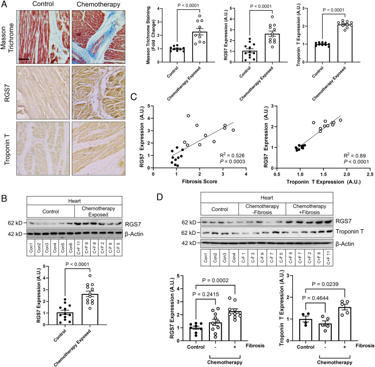
Fig. 1.
RGS7 is up-regulated in the chemotherapy-exposed myocardium. (A) Cardiac staining (Scale bar, 100 μm.) and (B) correlation analysis for RGS7, Troponin T, and Masson trichrome in control or chemotherapy-exposed patients (n = 10). (C) RGS7 expression in heart tissue from chemotherapy-exposed patients or controls (n = 12). (D) RGS7 (n = 8 to 10) and Troponin T (n = 4 to 5) immunoreactivity in control and chemotherapy patients with/without detectable fibrosis. Codes corresponding to specific chemotherapy patients and controls (SI Appendix, Table S2) are provided. β-Actin serves as a loading control for immunoblots. Exact P values are provided on graphs. Data are presented as mean ± SEM.

