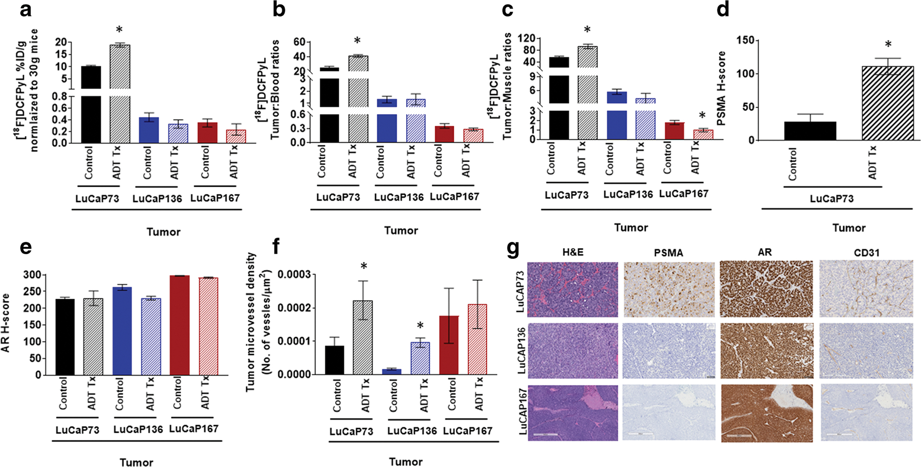
Fig. 5.

[18F]DCFPyL biodistribution (a), tumor to blood ratios (b; T:B), and tumor:muscle ratios (c; T:M) in control and ADT (degarelix) treated LuCaP73, LuCaP136, and LuCaP167 tumor-bearing mice. Biodistribution was performed at 60 min post [18F]DCFPyL injection. Each bar represents mean %ID/g ± SE (a), mean T:B± SE (b), or T:M ± SE (c); n = 5–7. (d)–(f) Quantitative histological analysis of PSMA (d, H-score), androgen receptor (e, AR, H-score), and CD31 (f, tumor microvessel density). Each bar in (d) and (e) represents mean H-score ± SD. Each bar in (f) represents the number of vessel/μm2 ± SD. (g) Representative H&E and IHC staining of tumor sections showing expression of PSMA, AR, and CD31; *significant difference in the control group versus the treated group (P < 0.05).
