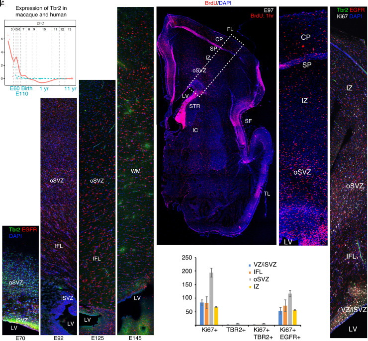
Fig. 3.
Cessation of neurogenesis and transition to gliogenesis by E97. (A) Transcriptome analysis using the online Psychencode database (34) (http://evolution.psychencode.org/#) indicates loss of Tbr2 expression in dorsolateral prefrontal cortex (DFC) prior to E110 in macaque (blue line) and prior to the equivalent developmental stage in humans (red line). Y-axis represents normalized gene expression as log2(RPKM+1) where RPKM is number of reads per million kilobases. The vertical gray lines separate developmental stages: 3, early fetal; 4 to 6, mid fetal; 7, late fetal; 8 to 9, infancy; 10 to 11, childhood; 12, adolescence; 13 to 15, adulthood. The vertical gray line between 7 and 8 represents birth in both species as described (34). (B–E) Immunohistochemistry (IHC) for Tbr2 and EGFR in coronal macaque dorsal parietal brain sections at the indicated ages. By IHC, robust Tbr2 expression was found in the iSVZ and oSVZ at E70 (B), but only a few cells were found at E92 (C). Tbr2 expression was not detected at E125 or E145 (D and E). In contrast, EGFR expression continues in the iSVZ and oSVZ through E145. (F) Acute BrdU (1 h) labeling shows the position of cells in S phase in a whole cerebral hemisphere; boxed region magnified in (G). (H) E97 IHC showing triple labeling of Ki67, Tbr2, and EGFR and quantified in (I). Images are confocal montages. (Scale bar, 200 μm in (B–E), (H), 800 μm in (F).) Error bars represent SEM. FL, frontal lobe; IC, internal capsule; LV, lateral ventricle; SF, Sylvian fissure; STR, striatum; TL, temporal lobe.

