Fig. 5.
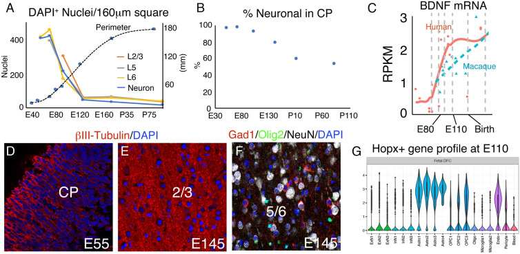
Neuropil growth, regulation, and cell-type diversification in the developing macaque cortical plate. (A) DAPI nuclear density in 160 μm square regions of single confocal images, by cortical layer as shown in Fig. 4A, with neuronal density plotted in blue. (B) Percentage of total DAPI-labeled nuclei that stain for neuronal markers; Tbr1 at E55, Tbr1/Ctip2/Brn2 at E70-E97, and NeuN at E145, P7, and P91. Neuronal density declines faster than cellular density after E70. (C) Single-cell transcriptome data via Psychencode showing that humans express higher levels of BDNF in dorsal frontal cortex than do macaques during gyrification. (D and E) Neuropil labeled by βIII-tubulin IHC. (F) Neurons, OPCs, and interneurons revealed by NeuN, Olig2, and Gad1, respectively. (G) Psychencode data showing that Hopx+ cells express gene clusters principally associated with astrocyte and OPC lineages at E110. [Scale bar: 40 μm in (D–F).]

