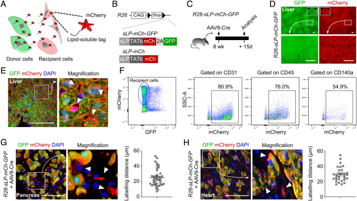
Fig. 1.
Generation of CILP for labeling of neighboring cells in vivo. (A) Cartoon showing the working principle of CILP. (B) Schematic showing generation of R26-sLP-mCh-GFP and R26-sLP-mCh mice lines. (C) Schematic showing the experimental design. (D) Whole-mount fluorescence images of livers from R26-sLP-mCh-GFP after AAV8-Cre treatment. (E) Immunostaining for GFP and mCherry on liver sections from R26-sLP-mCh-GFP after AAV8-Cre treatment. Arrowheads, GFP–mCherry+ cells. (F) FACS analysis of liver samples from R26-sLP-mCh-GFP after AAV8-Cre treatment. (G and H) Immunostaining for GFP and mCherry on sections of pancreas (G) or heart (H) samples from R26-sLP-mCh-GFP 3 wk after AAV-Cre treatment. Arrowheads, GFP–mCherry+ cells. Right panels showing the quantification of labeling distance. n = 40 cells for the pancreas, and n = 30 cells for the heart. Data are representative of 3 mouse samples. (Scale bars, 100 μm.)

