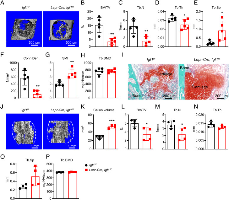
Fig. 3.
Deletion of Igf1 from BMSCs impairs bone regeneration after injuries. (A) Representative microCT images of the middle femur diaphysis of 12-wk-old male Lepr-Cre; Igf1f/f mice and littermate controls 7 d after bone drilling. (B–H) MicroCT analysis of trabecular bone volume ratio (B), trabecular number (C), trabecular thickness (D), trabecular spacing (E), connectivity density (F), SMI (G), and trabecular bone mineral density (H) in drilled bones (n = 5 mice per genotype from three independent experiments). (I) Representative safranin O/Fast Green images of the bone callus 14 d after mid-diaphyseal femur fracture. (J–P) MicroCT analysis of the bone callus 14 d after mid-diaphyseal femur fracture. Representative images (J), callus volume (K), trabecular bone volume ratio (L), trabecular number (M), trabecular thickness (N), trabecular spacing (O), and trabecular bone mineral density (P) were shown (n = 5 mice per genotype from three independent experiments). The statistical significance was assessed using two-tailed unpaired Student’s t test. Data represent mean ± SD (*P < 0.05, **P < 0.01).

