Fig. 7.
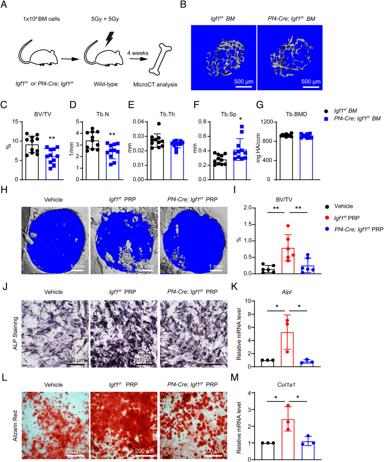
Deletion of Igf1 from MKs/PLTs reduces trabecular bones after BM transplantation and compromises the therapeutic effects of PRP. (A) Schematic illustration of the BM transplantation experiment. One million whole BM cells from Pf4-Cre; Igf1f/f mice or littermate controls were transplanted into lethally irradiated wild-type recipients. The bone phenotypes were analyzed 4 wk after transplantation by microCT. (B) Representative microCT images of the trabecular bone in the distal femur metaphysis. (C–G) MicroCT analysis of trabecular bone volume ratio (C), trabecular number (D), trabecular thickness (E), trabecular spacing (F), and trabecular bone mineral density (G) in recipient mice (n = 11 mice per genotype from two independent experiments). (H and I) In vivo treatment of the calvarial defects by PRP or vehicle control. Critical-size calvarial defects were treated with PRPs from Igf1f/f or Pf4-Cre; Igf1f/fmice, or vehicle control (saline), and analyzed 4 wk after treatment by microCT. Representative microCT images (H) and quantification of trabecular bone volume ratio (I) were shown (n = 6 mice per genotype from two independent experiments). (J–M) In vitro osteogenic differentiation of wild-type BMSCs after PRP or vehicle treatment. PRPs from Igf1f/f or Pf4-Cre; Igf1f/fmice, or vehicle control (PBS), were added to the osteogenic medium (1:50). Alkaline phosphatase (ALP) staining was performed 7 d after differentiation (J), and quantified by qPCR of Alpl (K). Alizarin red staining was performed 14 d after differentiation (L), and quantified by qPCR of Col1a1 (M) (n = 3 independent experiments). The statistical significance was assessed using two-tailed unpaired Student’s t test (C–G), or one-way ANOVA with Tukey's multiple comparisons test (I, K, M). Data represent mean ± SD (*P < 0.05, **P < 0.01).

