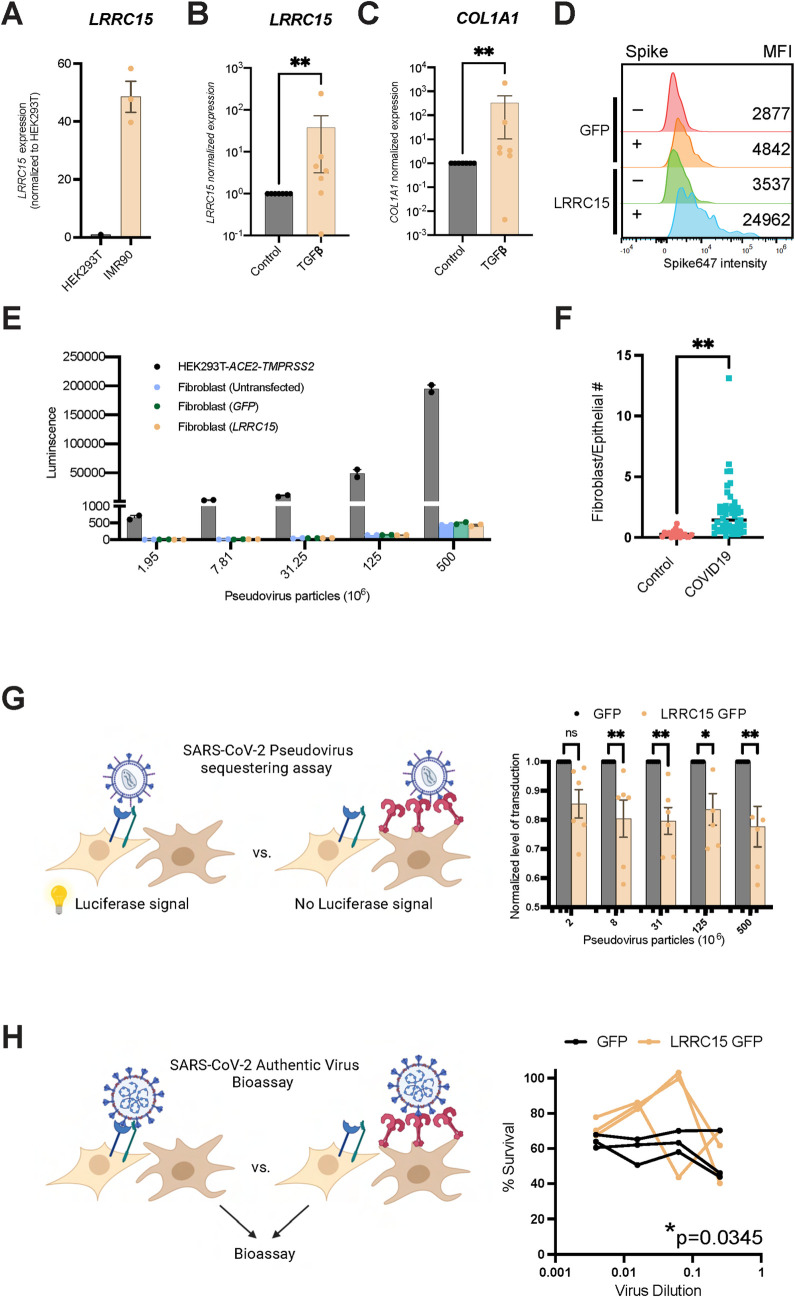
Fig 5. LRRC15 is not a SARS-CoV-2 entry receptor but inhibits infection in trans.
(A) IMR90 fibroblasts express LRRC15, quantified via RT-qPCR. N = 3 per cell line. (B and C) TGFβ increased (B) LRRC15 and (C) COL1A1 in fibroblasts, quantified via RT-qPCR. N = 7 for each group. Significance was determined by Mann–Whitney one-tailed test, **p < 0.01. (D) IMR90 fibroblasts expressing LRRC15 bind spike, MFI = mean fluorescence intensity. (E) Fibroblasts do not have innate tropism for SARS-CoV-2 and overexpression of LRRC15 does not mediate infection. Transduction efficiency (luciferase luminescence) was compared to permissive cell line HEK293T-ACE2-TMPRSS2. N = 2 independent replicates for each group. (F) Pooled analysis of 3 independent studies indicate ratio of fibroblasts to epithelial cells in COVID-19 lungs is approx. 2:1 (0.3 in control (n = 19) and 2.06 in COVID-19 (n = 47); unpaired two-tailed t test, p < 0.0001). (G) LRRC15 expressing fibroblasts can suppress SARS-CoV-2 spike pseudovirus infection of HEK293T-ACE2-TMPRSS2 cells. Significance was determined by two-way ANOVA, Sidak’s multiple comparison test, **p < 0.01,*p < 0.05. N = 6 per condition. (H) LRRC15 expressing fibroblasts can suppress authentic SARS-CoV-2 infection of HEK293T-ACE2-TMPRSS2 cells. Significance was determined by two-way ANOVA, Sidak’s multiple comparison test, *p < 0.05. N = 3 per condition. The data underlying all panels in this figure can be found in DOI: 10.5281/zenodo.7416876. ACE2, angiotensin-converting enzyme 2; COL1A1, collagen type I alpha 1 chain; LRRC15, leucine-rich repeat-containing protein 15; SARS‑CoV‑2, Severe Acute Respiratory Syndrome Coronavirus 2; TMPRRS2, Transmembrane serine protease 2.

