. 2023 Jan 25;12:e84477. doi: 10.7554/eLife.84477

Figure 1. SARS-CoV-2 Orf3a colocalizes with markers for the plasma membrane and the endocytic pathway by live-cell imaging.

(A) Summary table of SARS-CoV-2 (CoV-2) Orf3aHALO colocalization with subcellular protein markers. All markers used to identify cellular compartments are listed in the table in A and are transiently expressed (mEm, mEmerald; mNG, mNeonGreen; GFP, green fluorescent protein). (B) Live-cell image of transiently expressed farnesylated-GFP (green) and CoV-2 Orf3aHALO (magenta) using a HEK293 doxycycline-inducible CoV-2 Orf3aHALO stable cell line. White arrows indicate co-localization. (C) Total Internal Reflection Fluorescence (TIRF) imaging of HEK293 cell with transient expression of CoV-2 Orf3aHALO (white). Orange box, magnification of the surface to highlight CoV-2 Orf3aHALO (black). (D–I) Live-cell image of transiently expressed (D) Rab5-mEm, (E) Rab7-mEm, (F) LAMP1-mEm, (G) Sec61-mEm, (H) αMannosidase-II-mEm, or (I) Pex11-mNG (green) with CoV-2 Orf3aHALO (magenta) as described in (B). White boxes indicate regions of co-localization. All confocal images are representative of three to six independent experiments.

Figure 1.

Figure 1—figure supplement 1. SARS-CoV-2 Orf3a colocalizes with markers of the endocytic pathway, but not with a Golgi marker, by immunostaining.

Figure 1—figure supplement 1.

(A) Summary table of SARS-CoV-2 (CoV-2) Orf3aHALO colocalization with subcellular antibody markers. All markers used to identify cellular compartments are listed in the table in A. (B) Fixed HEK293 cells without (Dox -) and with (Dox +) doxycycline to induce expression of CoV-2 Orf3aHALO (magenta). Cell nuclei are visualized with Hoechst 33342 stain (blue). (C–F) Fixed HEK293 cells treated with doxycycline to induce expression of CoV-2 Orf3aHALO (magenta) stained with (C) EEA1, (D) Rab7, (E) LAMP1, and (F) TGN46 (green). Nuclei are labeled with Hoechst 33342 (blue). All images are representative of three independent experiments.
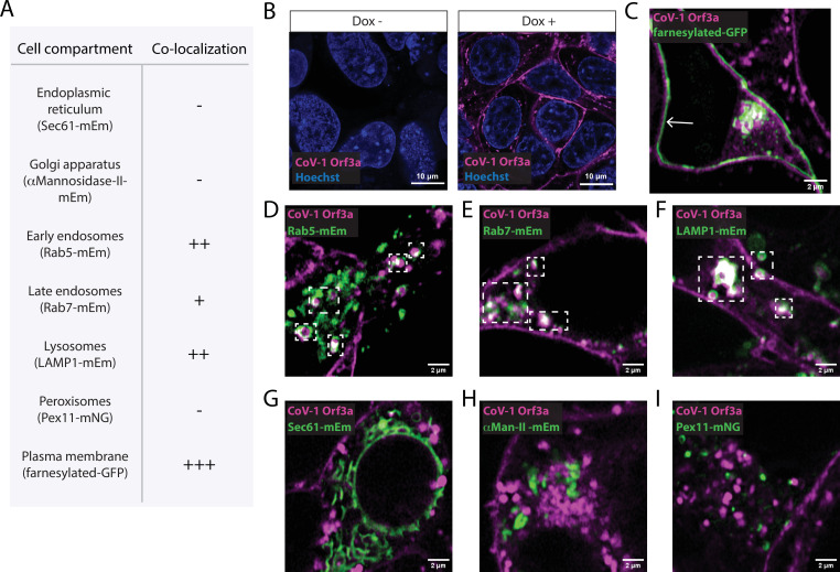
Figure 1—figure supplement 2. SARS-CoV-1 Orf3a colocalizes with markers for the plasma membrane and the endocytic pathway by live-cell imaging.

Figure 1—figure supplement 2.

(A) Summary table of SARS-CoV-1 (CoV-1) Orf3aHALO colocalization with subcellular protein markers. All markers used to identify cellular compartments are listed in the table (A) and are transiently expressed (mEm, mEmerald; mNG, mNeonGreen; GFP, green fluorescent protein). (B) Live-cell imaging of HEK293 cells without (Dox -) and with (Dox +) doxycycline to induce expression of CoV-1 Orf3aHALO (magenta). Cell nuclei are visualized with Hoechst 33342 stain. (C–I) Live-cell image of transiently expressed (C) farnesylated-GFP, (D) Rab5-mEm, (E) Rab7-mEm, (F) LAMP1-mEm, (G) Sec61-mEm (H) αMannosidase-II-mEm or (I) Pex11-mNG (green) with CoV-1 Orf3aHALO (magenta) using the HEK293 stable cell line in (B). White arrows and boxes indicate regions of co-localization. All images are representative of three independent experiments.