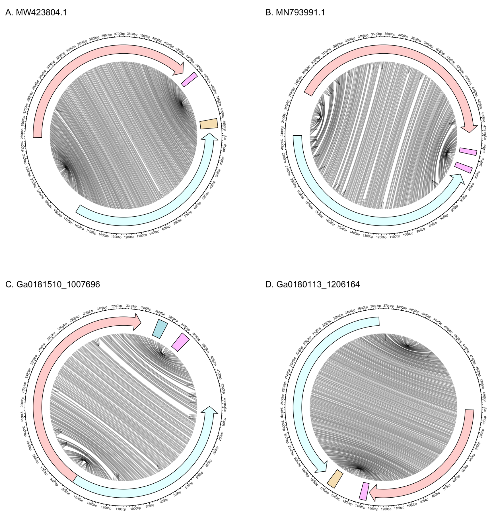
Figure 4:

Genomic and secondary structure of Armillaria borealis ambi-like virus 1 (A), Tulasnella ambivirus 1 (B), and ambi-like sequences discovered here (C, D). Red and blue denote (+) and (−) polarities, respectively. Lines connect bases in the genome that are paired in the predicted secondary structure. Arrows represent ORFs and rectangles represent self-cleaving ribozymes. The (+) and (−) ribozymes are HHR3 and HPR-meta1 in (A), HHR3 and HHR3 in (B), CPEB3 and HHR3 in (C), and HHR3 and HPR-meta1 in (D). In all cases, the ribozymes are located outside the ORFs at the end of the rod.
