Fig. 3.

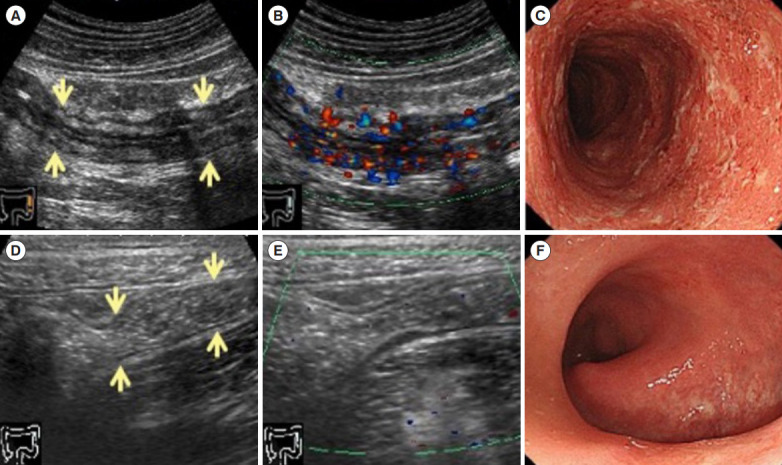
Ultrasonography and colonoscopy findings of immune-mediated colitis (IMC) in a patient who had anti-PD-L1 antibody therapy (durvalumab). IMC was resistant to steroids and anti-tumor necrosis factor α antibodies. The remission was inducted using vedolizumab. At the onset of colitis, ultrasonography (US) shows a thickened bowel wall, the stratified structure is partially indistinct (arrows) (A), and color Doppler shows an increased blood flow signal (B). Colonoscopy (CS) shows a circular erosion from the cecum to the rectum, and there are exudates and spontaneous bleeding (C). After administering vedolizumab, US shows the bowel wall thickening improvement, and the stratified structure becomes clearly visible (arrows) (D), color Doppler shows no blood flow signal (E). CS findings improves that the erosion and exudates are disappeared, and the mucosal vascular pattern becomes also visible (F). PD-L1, programmed cell death receptor ligand 1.
