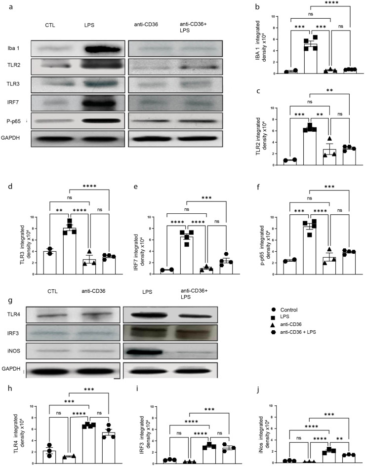
Figure 4.
Blocking CD36 significantly alters TLR-2 and TLR-3 signalling. (a) Western blot of whole brain protein lysates from the control (saline), anti-CD36, LPS and anti-CD36 + LPS injected pups. (b) Iba1, (c) TLR2, (d) TLR3, (e) IRF7, (f) phosrpho-P65 expression levels were quantified in different conditions. GAPDH was used as loading control A significant increase in the levels of all the tested proteins is observed after LPS injection. Treating the pups with anti-CD36 before LPS injections considerably reduces their expression to the level as observed in the control pups. (g) Western blot of total protein lysates from the control, anti-CD36, LPS and anti-CD36 + LPS injected mice. (h) TLR4, (i) IRF3, (j) iNOS expression levels were quantified in above-described conditions. GAPDH is used as loading control. A significant increase in the levels of all the proteins tested is observed after LPS injection when compared to control and anti-CD36 treated mice. Of note, LPS-mediated increase in expression of TLR4/IRF3 is maintained in the anti-CD36 + LPS treated group. The entire data was presented as mean ± SEM and statistical significance between the groups was achieved using one-way ANOVA with Tukey’s multiple comparison test (ctl vs LPS, ctl vs anti-CD36, LPS vs anti-CD36 + LPS) and depicted as ****p ≤ 0.0001; ***p ≤ 0.001; **p ≤ 0.01 and *p ≤ 0.05.

