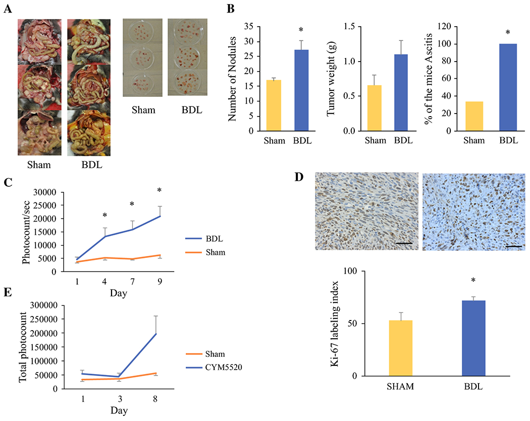
Figure 7.

Peritoneal carcinomatosis model results. a. Post-sacrifice comparison of carcinomatosis and peritoneal nodules on day 14 after intraperitoneal injection. Jaundice is clearly visualized in the BDL group compared to sham laparotomy (left). BDL was associated with increased number and size of peritoneal nodules (right). b. Numbers of nodules, total weights of all nodules and percentage of the mice with ascites after BDL compared to sham laparotomy. c. In vivo BLI demonstrates increased tumor burden in mice following BDL. d. Paraffin-embedded tumor sections were immunostained with Ki-67 (top). Ki-67 labeling index is increased in mice following BDL (bottom). e. Effect of TCAs in BDL is mimicked by S1PR2 agonist CYM5520. Data are expressed as mean±SEM, n=5, *, P<0.05, compared with the control group.
