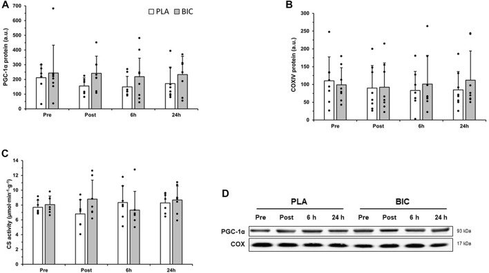
FIGURE 5.
Comparison of (A) PGC-1α, and (B) COXIV protein abundance in the vastus lateralis between the PLA (white bars) and BIC (grey bars) conditions. Values are normalized to the α-tubulin loading control and expressed in arbitrary units. All values are mean ± SD (n = 8). Individual data is shown in black points. (C) Comparison of citrate synthase (CS) activity between the PLA (white bars) and BIC (grey bars) conditions. Values are mean ± SD (n = 8). (D) Representative western blots for each protein.

