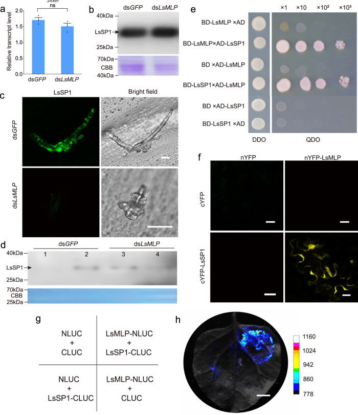
Fig. 3. LsSP1 binds to a salivary sheath protein LsMLP.
a, b Influence of dsLsMLP treatment on the LsSP1 abundance in salivary gland quantified by qRT-PCR (a) and western-blotting (b) assays. Data are presented as mean values ±SEM (n = 3 independent biological replicates). P-values were determined by two-tailed unpaired Student’s t test. ns, not significant. c LsSP1 staining of salivary sheath secreted from dsRNA-treated Laodelphax striatellus. The salivary sheaths secreted from dsGFP- or dsLsMLP-treated L. striatellus were probed with the anti-LsSP1 serum conjugated with Alexa Fluor™ 488 NHS Ester (green), and examined by Leica SP8. Bars = 20 μm. d Detection of LsSP1 in watery (lane1, lane3) and gel saliva (lane 2, lane 4) secreted from dsGFP- or dsLsMLP-treated L. striatellus. e Yeast two hybrid assays showing the interaction between LsSP1 and LsMLP. The different combinations of constructs transformed into yeast cells were grown on the selective medium SD/-Trp/-Leu (DDO), and the interactions were tested with SD/-Trp/-Leu/-His/-Ade (QDO). f Bimolecular fluorescence complementation (BiFC) assays showing the interaction between LsSP1 and LsMLP. Bars = 20 μm. g The co-expression scheme in Nicotiana benthamiana leaves during Luciferase complementation (LUC) assays. h Results from LUC assays showing the interaction between LsSP1 and LsMLP. Bar = 1 cm. Experiments in b, c, d, f, and h were repeated three times with the similar results. Source data are provided as a Source Data file.

