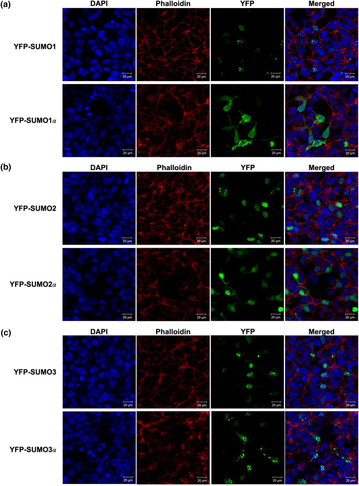
Figure 7.
The SUMO alphas exhibit different cellular localization patterns from those associated with their prototypical isoform. HEK293A cells were transfected with expression constructs coding for YFP-fusion forms of either the prototypical SUMO proteins or their associated SUMO alpha isoforms. At 24 h post-transfection, the cells were fixed, permeabilized, treated with Phalloidin (a bicyclic peptide that stains all types of actin filaments) and DAPI (a DNA-binding fluorescent dye), and images were captured by confocal microscopy. The images provided constitute representative fields. (a) YFP-SUMO1 and YFP- SUMO1α. (b) YFP-SUMO2 and YFP- SUMO2α. (c) YFP-SUMO3 and YFP- SUMO3α. The fluorescence associated to each marker and its respective color representation are as follows: DAPI, blue; Phalloidin, red; YFP, green.

