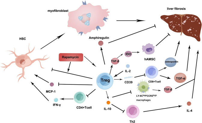
Fig. 2. The role of Treg in inhibiting fibrosis.
IL-2 as well as its complexes accelerate Tregs expressing CD39 in the liver, thereby inhibiting the multiplication of CD8+ T cells and its function of generating TNF-α and osteopontin, which can reduce biliary fibrosis. Tregs can regulate the pro-fibrotic roles of Th2 cells and Ly-6ChighCCR2high inflammatory monocytes/macrophages that secrete IL-4 and TGF-β respectively, interestingly, IL-10 secreted by Tregs may regulate them. Tregs can regulate the TGF-β-IDO signalling pathway to enhance the function of hAMSC that repress liver fibrosis. Tregs promoting the expression of amphiregulin can inhibit the development of fibrosis by promoting the proliferation of hepatocytes. In CCL4-induced liver fibrosis, rapamycin has an effective protective effect on the liver. Rapamycin significantly increased the functional activity of CD4+CD25+ Tregs and enhanced the inhibitory ability of Tregs on HSCs activation. Tregs can repress MCP-1 which plays an important role in liver fibrosis by activating HSC production. Tregs can directly repress CD4+ T cell expression that reduces IFN-γ activated HSCs. Tregs also directly promote amphiregulin to repress liver fibrosis. Footnote: interleukin (IL), regulatory cells (Treg), transforming growth factor-α (TGF-α), T helper cell (Th), indoleamine 2,3-dioxygenase (IDO), human amniotic mesenchymal stromal cell (hAMSC), carbon tetrachloride(CCL4), hepatic stellate cells (HSCs), monocyte chemotactic protein-1 (MCP-1), Interferon-γ (IFN-γ).

