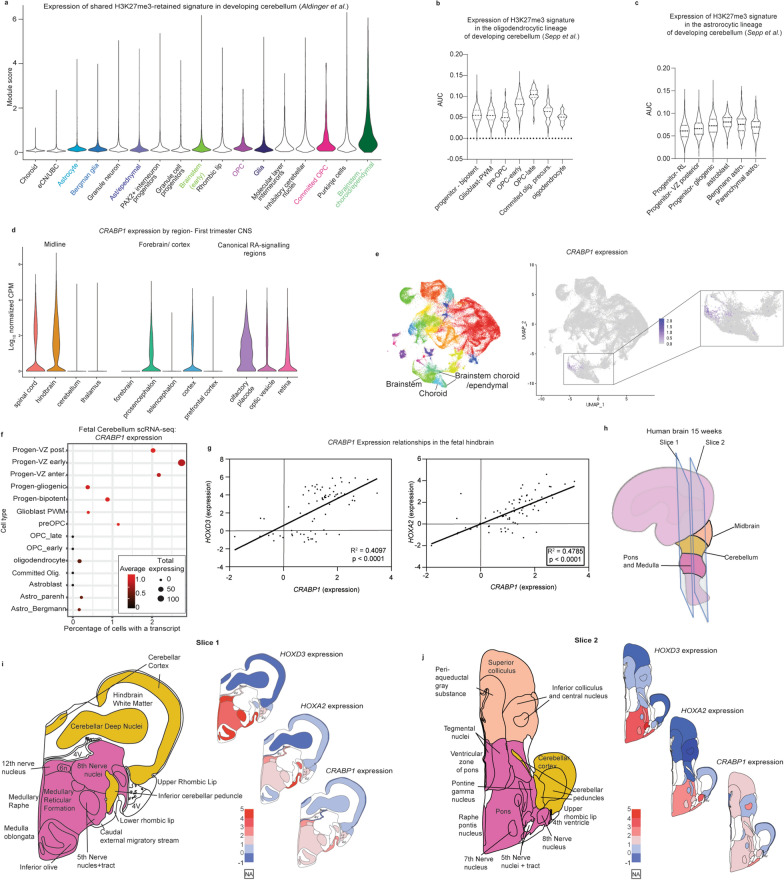
Fig. 6.
Common H3K27me3-retained signature mirrors human hindbrain brain developmental patterns. a Expression of the shared DMG/PFA-derived H3K27me3 gene signature in scRNA-seq data of the developing human posterior fossa (Aldinger et al.) [46] grouped by cell type. Highlighted groups indicate cell types pertinent to DMGs/PFAs. b H3K27me3 gene-signature expression along the oligodendrocyte lineage in an independent scRNA-seq atlas of the developing posterior fossa (Sepp et al., unpublished) [48]. c H3K27me3 gene-signature expression along the astrocytic lineage in the scRNA-seq atlas of the developing posterior fossa used in b. d First-trimester human brain scRNA-seq [47] expression patterns of CRABP1 grouped by anatomic region. Left: midline structures, middle: forebrain and cortical structures, right: regions with canonical retinoic acid (RA) signaling. e CRABP1 expression patterns in the developing cerebellum scRNA-seq atlas from a. Left: UMAP embedding colored by cluster, middle: CRABP1 expression across all cells in UMAP embedding, right (inset): magnified depiction of CRABP1 expression patterns in a subset of cells including brainstem and brainstem-derived choroidal/ependymal cells. f CRABP1 expression patterns in astrocytic and oligodendrocyte lineages of the developing cerebellum scRNA-seq atlas from b and c. Early progenitors are grouped along the top of the Y-axis. The more differentiated oligodendrocyte and the astrocytic lineages are each grouped below along the Y-axis. The percentage of cells expressing at least one CRABP1 transcript (X-axis), the total number of cells expressing at least one CRABP1 transcript (point size) and the average expression value (color) are depicted. g Microarray-based log2 expression levels from micro-dissected fetal hindbrain tissue at 15 and 16 weeks post-conception from BrainSpan’s prenatal lateral microdissection (LMD) Microarray comparing CRABP1 expression patterns to those of HOXA2 and HOXD3. Data were analyzed with a simple linear regression and R-squared calculated with a goodness of fit test with 95% confidence intervals. h Schematic depicting sectioning of the 15–16 weeks post conception fetal brain utilized for depictions in g, I, and j. Early anatomic structures of the infratentorial brain are colored in peach (midbrain), yellow (cerebellum), and deep pink (pons and medulla). i-j Expression of HOXD3, HOXA2, and CRABP1 at 15 and 16 weeks post conception mapped to infratentorial brain at slices 1 (i) and slices 2 (j) from h. Left: schematic of brain sub-structures colored by overarching structure corresponding to key in h. Right, from top to bottom heatmaps of HOXD3, HOXA2, and CRABP1 expression in corresponding areas. Illustration adapted from reference figure from the Allen Brain Atlas

