Fig. 5.
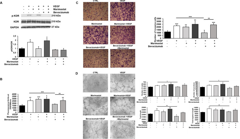
Effects of VEGF stimulation and its inhibition by Bevacizumab on ECFC proliferation, invasion and tubular structure formation. A Western blotting results show the phosphorylation of KDR in ECFCs after VEGF stimulation (50 ng/ml) and its inhibition by Bevacizumab (5 µg/ml) treatment in mesenchymal and amoeboid conditions. Numbers on the right refer to molecular weights expressed in kDa. Histograms report band densitometry. Results are the mean of 5 different experiments performed in duplicate, and are shown as mean value ± SD. B The effects of VEGF stimulation and its inhibition were also observed on cell proliferation (B), cell invasion (C) and in vitro angiogenesis (D). Cell proliferation was quantified by AlamarBlue® assay and fluorescence was measured at 530 nm/590 nm. n = 3 independent samples. Boyden chamber invasion assay and capillary morphogenesis were performed adding Marimastat to the Matrigel solution before polymerization and VEGF (25 ng/ml) ± Bevacizumab (5 µg/ml) in cell suspension. Representative microphotographs (×10) of migrated cell filters and capillary-like structures are shown. Histograms in C refer to quantification of Matrigel invasion assay obtained by counting the total number of migrated cells/filter. Capillary network was quantified by Angiogenesis Analyzer Image J tool. Histograms in D represent the mean number of nodes, number of junctions, total length, and total length of segments respectively. Data are representative of measures obtained from at least nine fields. Results are the mean of 5 different experiments performed in duplicate and are shown as mean value ± SD. *p < 0.05; **p < 0.001; ***p < 0.0001 significantly different from control or the experimental point indicated

