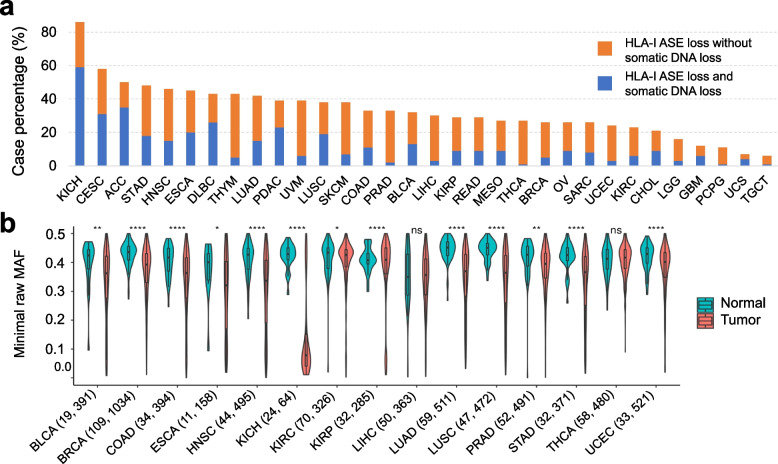
Fig. 2.
Pervasiveness of HLA-I allele-specific expression loss. a Proportions of HLA-I ASE loss across TCGA subtypes (orange bars) as inferred using arcasHLA-quant. Blue (orange) bars represent proportions of cases where expression loss is (not) accompanied by somatic DNA loss, as inferred by LOHHLA on WES data. b HLA-I ASE comparison between tumor and normal cases in TCGA cohorts. HLA-I ASE is captured by the minimal raw minor allele frequency among the three HLA-I genes (minimal raw MAF). Numbers in the parentheses indicate the normal and tumor case numbers respectively. Only the TCGA cohorts with more than 10 normal cases are shown. Significance labels: “ns” or nothing labeled: p > 0.05; “*”: p < 0.05; “**”: p < 0.01; “***”: p < 0.001; “****”: p < 0.0001

