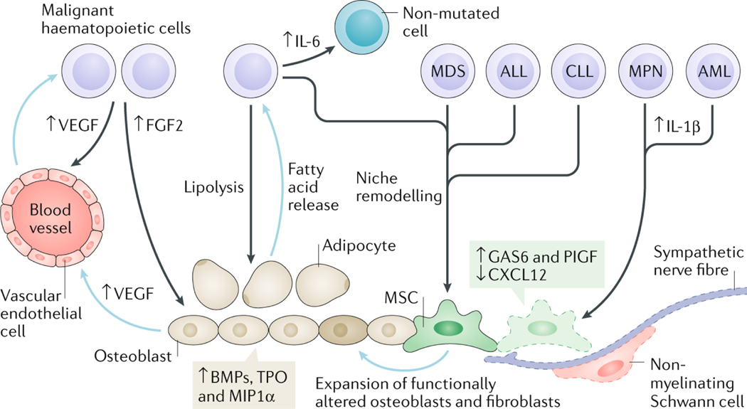
Fig. 2: Bone marrow niche remodelling favours disease progression in haematological malignancies.
Representative features of the remodelled bone marrow (BM) niche contributing to the progression of different haematological malignancies. These diseases can arise through genetic and/or epigenetic alterations in haematopoietic cells causing remodelling of the niche that supports their own growth and survival at the expense of normal haematopoiesis. Myeloproliferative neoplasm (MPN) and acute myeloid leukaemia (AML) cells cause neuroglial damage (affecting Schwann cells and sympathetic nerve fibres) in the BM and reduced expression of CXC-chemokine ligand 12 (CXCL12) in mesenchymal stem cells (MSCs). In MPN, the neuroglial damage is caused by increased production of cytokines, like interleukin-1β (IL-1β), leading to apoptosis of Nestin-GFP+ MSCs, which can be rescued through chronic treatment with sympathicomimetic drugs that indirectly improve reticulin fibrosis in mice and humans. In AML, a reduction in BM sympathetic innervation has been correlated with proliferation of Nestin-GFP+ MSCs primed for osteoblastic differentiation at the expense of neural–glial antigen 2 (NG2)+ periarteriolar niche cells, although the potential relevance of these alterations in human AML is unclear. Increased growth arrest-specific 6 (GAS6) and placental growth factor (PlGF) expression by BM MSCs fosters leukaemia cell survival, proliferation and therapy resistance. Vascular endothelial growth factor (VEGF) secreted by both malignant haematopoietic cells and osteoblasts stimulates angiogenesis. In addition, other factors produced by endothelial cells (such as granulocyte–macrophage colony-stimulating factor (GM-CSF), IL-6 and stem cell factor (SCF)) promote survival and proliferation of malignant haematopoietic cells. IL-6 produced by chronic myeloid leukaemia (CML) cells renders non-mutated haematopoietic and stromal cells pro-inflammatory. Malignant haematopoietic cells stimulate expansion of stromal cells through secretion of growth factors, such as bone morphogenetic proteins (BMPs). A reciprocal relationship occurs between malignant haematopoietic cells and adipocytes wherein malignant cells induce lipolysis from adipocytes and, in turn, adipocytes release fatty acids, which are used as an energy source by malignant haematopoietic cells. Blue arrows in this figure indicate processes or factors secreted by niche cells and black arrows indicate processes or factors released by malignant haematopoietic cells. ALL, acute lymphoblastic leukaemia; CLL, chronic lymphocytic leukaemia; FGF2, fibroblast growth factor 2; MDS, myelodysplastic syndrome; MIP1α, macrophage inhibitory protein 1α; TPO, thrombopoietin.

