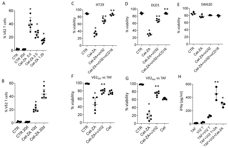
Figure 4.
Cet-ZA can expand anti-tumor Vδ2 T cells and stimulate cytolytic activity. (A): Purified T lymphocytes from healthy donors (#49, #50) were co-cultured with CRC-TAF 16-001, or 16-004 or 16-035 without (CTR) or with Cet-ZA (5 µg/mL, 2.5 µg/mL or 1.25 µg/mL) and IL-2 and analyzed on day 10 by flow cytometry. Results are expressed as percentage Vδ2 T lymphocytes; the mean ± SD from 6 experiments is also shown. (B): Experiments performed as in A without (CTR) or with Cet-ZA at 2.5 µg/mL for 10 or 20 days; results expressed as percentage of Vδ2 T lymphocytes also showing the mean ± SD from 6 experiments from co-cultures of T (#51, #52 and CRC-TAF (16-001 or 16-004 or 16-035). (C–E): Activated Vδ2 T cells obtained from healthy donors (n = 5) were used as effectors in cytotoxicity assay vs. HT29 or DLD1 ((C,D), EGFR+) or SW620 (E, EGFRdull) without (CTR) or with Cet-ZA (2.5 µg/mL) or Cet (2.5 µg/mL) or soluble ZA (1.2 µM). In some samples the anti-Vδ2 TCR mAb or the anti-CD16 mAb were added (1 µg/mL). Cytotoxicity was detected by crystal-violet staining and expressed as percent of viability. (F,G): Activated Vδ2 T lymphocytes obtained with ZA-activated CRC-TAF (Vδ2 TAF) or monocytes (Vδ2Mo) were employed as effectors against CRC-TAF as above. Cytotoxicity was detected and expressed as in (C–E). (H): IFNγ measured in the supernatant of Vδ2 T cells (#50) either alone or co-cultured with CRC-TAF (16-001 or 16-030 or 16-035), without or with 1.25 µM ZA or 2.5 µg/mL Cet-ZA, by ELISA. Results are expressed as pg/mL referred to a standard curve. In (A–G) panels: * p < 0.001 vs. CTR, ** p < 0.001 vs. Cet-ZA. In (H) panel: ** p < 0.004 vs. TAF + Vδ2 T and * p < 0.001 vs. TAF + Vδ2 T.

