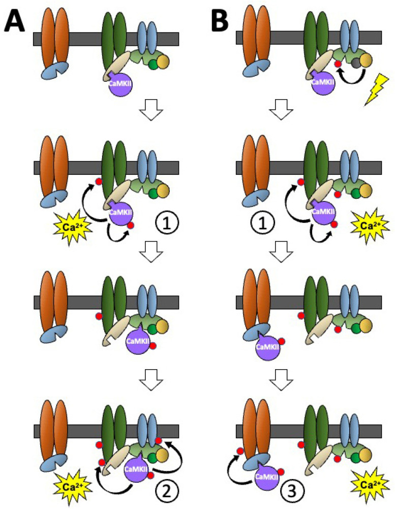
Figure 4.
A schematic diagram showing how stimulus-induced translocation of CaMKII between different binding proteins in a molecular environment can produce different functional responses to the activation of CaMKII by two sequential stimuli of the same type and how the influence of another signalling pathway can further modify the CaMKII response. The effects of two sequential stimulus-induced rises in calcium on non-phosphorylated CaMKII, when the surrounding binding partners are (A) also non-phosphorylated, or (B) phosphorylated. Coloured oblong shapes represent generic proteins with which CaMKII (purple disc) binds directly or indirectly. Dark grey bar represents a membrane. Geometric protrusions from CaMKII, and indents in the binding proteins represent binding sites for CaMKII. The small red disc represents the phosphorylation of a protein. See text for explanation of the sequence of events shown in the Figure.

