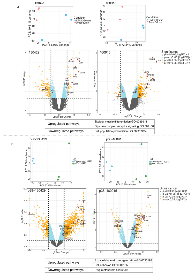
Figure 1.
Distinct set of genes and pathways upregulated in the anisomycin-treated 130429/160915 cells and p38–130429/160915 cells. (A): Principal component analysis of 30 min anisomycin-treated 130429/160915 cells compared with DMSO-treated 130429/160915 cells (normalized + log2). Volcano plot showing differentially expressed genes (DEGs) in 30 min anisomycin-treated 130429/160915 cells compared with DMSO-treated 130429/160915 cells (FDR threshold: 0.05). The most upregulated and downregulated pathway as indicated by the over-representation analysis (ORA), p-value < 0.05. (B): Principal component analysis of p38–130429/160915 cells compared with EV control–130429/160915 cells (normalized + log2). Volcano plot showing differentially expressed genes (DEGs) in p38–130429/160915 cells compared with EV control–130429/160915 cells (FDR threshold: 0.05). Most upregulated pathway as indicated by over-representation analysis (ORA) and most downregulated pathway as indicated by KEGG, p-value < 0.05.

