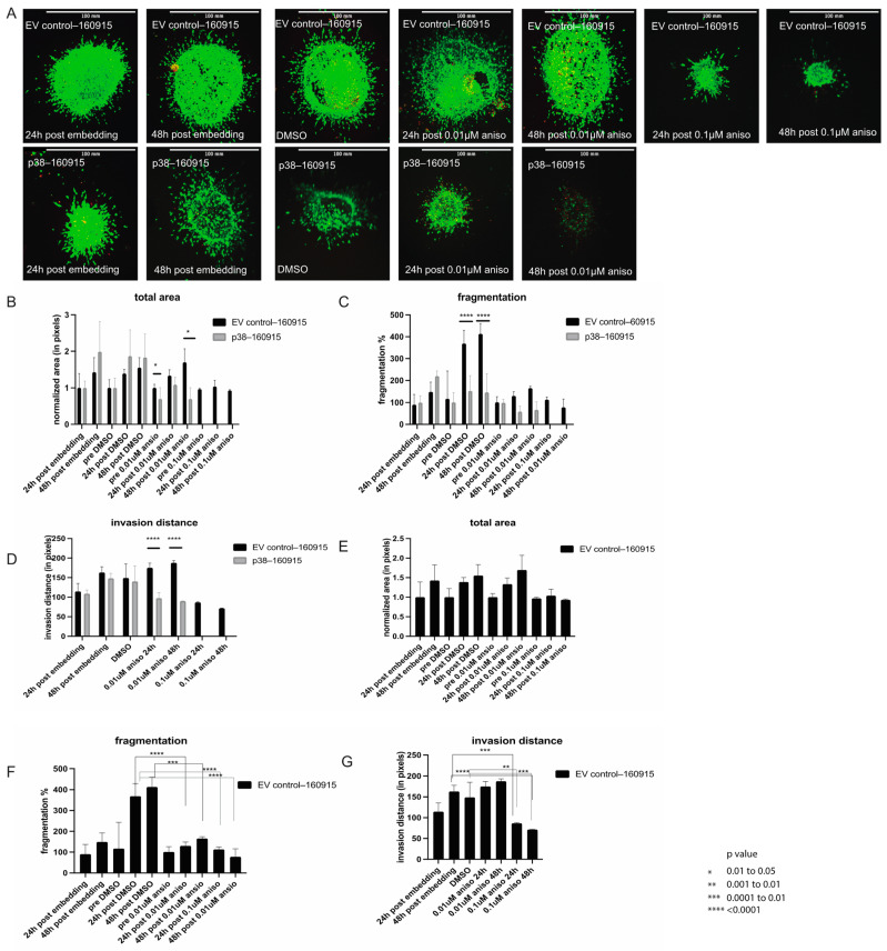
Figure 2.
Invasion assay in spheroids and protein expression of MITF. (A): EV control–160915 and p38–160915 spheroids were stained with calcein and ethidium and compared at 24, 48 and 72 h post-embedding in collagen with and without anisomycin/DMSO. (B): Total area was compared at 24, 48 and 72 h post-embedding in collagen in addition to comparing the total area between DMSO, 0.01 µM, and 0.1 µM anisomycin treatments lasting 24 and 48 h in both EV control–160915 and p38–160915. A significant difference was observed between EV control–160915 and p38–160915 at 24 h post embedding in collagen and prior to anisomycin treatment (p = 0.0138). as well as 72 h post-embedding in collagen and 48 h post 0.01 µM anisomycin treatment (p = 0.0197). Bars labelled as pre-DMSO/aniso refer to the spheres before the respective treatments. (C): Fragmentation was compared at 24, 48, and 72 h post-embedding in collagen in addition to a comparison of the fragmentation between DMSO, 0.01 µM, and 0.1 µM anisomycin treatments lasting 24 and 48 h in both EV control–160915 and p38–160915. A significant difference was observed between EV control–160915 and p38–160915 24 and 48 h post-DMSO treatment (p < 0.0001). Bars labelled as pre-DMSO/aniso refer to the spheres before the respective treatments. (D): Invasion distance was compared at 24, 48, and 72 h post-embedding in collagen in addition to a comparison of the invasion distance between DMSO, 0.01 µM, and 0.1 µM anisomycin treatments lasting 24 and 48 h in both EV control–160915 and p38–160915. A significant difference was observed between EV control–160915 and p38–160915 24 and 48 h post-0.01 µM anisomycin treatment (p < 0.0001). Invasion distance was calculated using images from the confocal microscope. (E): Total area was calculated using the same methods as in B except that the comparison group is limited to EV control–160915 only. (F): Fragmentation was calculated using the same methods as in C except that the comparison group is limited to EV control–160915 only. A significant difference was observed between 24 h DMSO-treated spheroids and 0.01 µM anisomycin treated spheres at 24 and 48 h (p < 0.0001). (G): Invasion distance was calculated using the same methods as in D except that the comparison group is limited to EV control–160915 only. A significant difference was observed between 48 h post-embedding in collagen and 0.1 µM anisomycin treatment at 24 (p < 0.002) and 48 h (p < 0.0001). A significant difference was observed between DMSO-treated spheroids and 0.1 µM anisomycin-treated spheroids at 24 (p < 0.0027) and 48 h (p < 0.001). This is a representative picture of one replicate out of the three biological replicates. The Experiment was repeated at least three times with three biological replicates.

