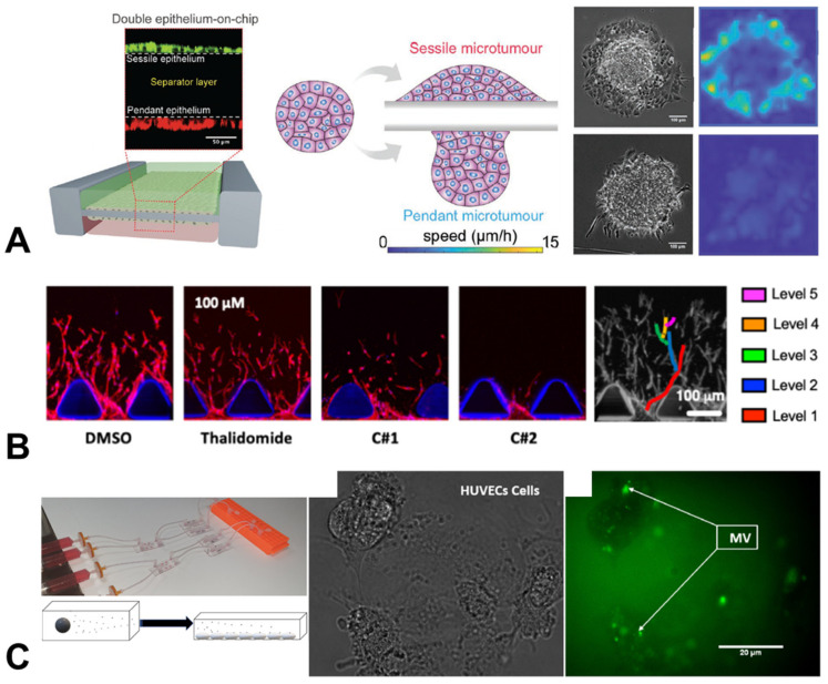
Figure 4.
Organ-chip models of cancer-associated behaviours. (A) The spreading speed of liver microtumour spheroids depended on the epithelial layer on which they are placed (scale bar = 100 µm) [44]. (B) Angiogenic sprouting, modelled and analysed in a microfluidic chip under a range of different anti-angiogenic drug treatments (thalidomide, C#1, C#2) (scale bar = 100 µm) [45]. (C) Chips placed in series allowed the monitoring of the uptake of procoagulant microvesicles secreted by glioblastoma and ovarian carcinoma cells (MV, green) by human endothelial cells (scale bar = 200 µm) [46]. Figures reproduced with permission.

