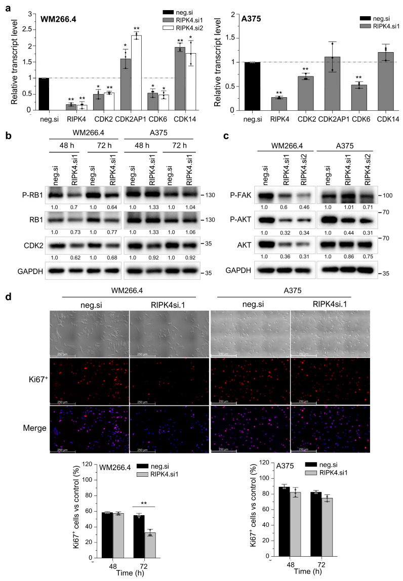
Figure 7.
Downregulation of RIPK4 alters proliferation in WM266.4, but not in A375 cells. WM266.4 and A375 cells were transfected with RIPK4.si1, RIPK4.si2, or neg.si RNA and analyzed, n = 3. (a) Transcript levels of RIPK4, CDK2, CDK6, CDK14CDK2AP1 48 h after transfection normalized to GAPDH. (b,c) Western blot images showing the levels of (b) proteins P-RB1, RB1, and CDK2, (c) proteins P-FAK, p-AKT, and AKT proteins in A375 and WM266.4 cells. GAPDH is the loading control. (d) Phase-contrast images of cells and immunofluorescence images of Ki67 protein (red) and cell nuclei (blue) 72 h after transfection. Original magnification: 100×, scale bar = 250 µm. Each bar represents the mean ± SD of three biological replicates. All RIPK4.si transfected cells were compared with neg.si (scrambled control) samples. Statistical analysis was performed using the Student’s t test. * p < 0.05 and ** p < 0.001 were considered significant.

