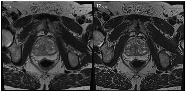
Figure 3.
Sixty-nine-year-old man with suspicion of prostate cancer. Deep learning image reconstructed thin-slice imaging (T2DLR) is shown on the right-hand side and standard imaging (T2S) on the left-hand side. T2DLR shows improved levels of sharpness and improved delineation of the prostate parenchyma. Biopsy revealed no malignancy.

