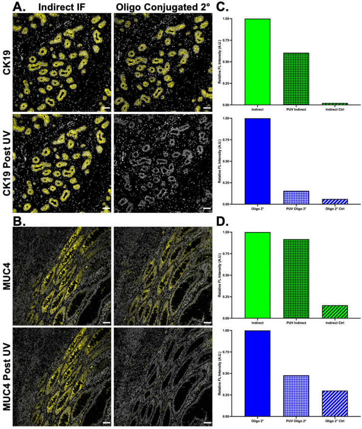
Figure 6.
Oligonucleotide conjugation to secondary antibodies. Secondary antibodies were conjugated to unique DS and used to stain (A) CK19 (yellow) and (B) MUC-4 (yellow) in tissues with DAPI (white) staining to show tissue patterns. The oligonucleotide-conjugated secondary antibody staining pattern was compared to indirect IF. Following imaging, both samples were treated with UV light, where the oligonucleotide-conjugated secondary antibodies showed substantially decreased staining. The staining intensity (green) and signal removal (blue) were quantified for both (C) CK19 and (D) MUC4. Images shown are representative of multiple fields of view and quantification results are reported as a representative average of all images. Scale bar = 50 μm.

