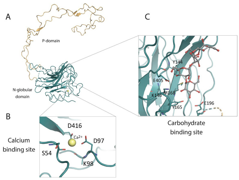
Figure 4.
Crystal structure of the calnexin luminal domain and its characteristics. (A) Crystal structure of the calnexin intraluminal domain. The globular N-globular domain is shown in green while the P-domain is depicted in yellow. (B) Calnexin putative Ca2+-binding site, showing a Ca2+ ion (yellow circle) coordinated by Asp416, Asp97, Ser54 and potentially Lys98. (C) Calnexin carbohydrate binding site, showing the sidechains of Tyr144, Lys146, Tyr165, Glu196, Glu405 and Met168 involved in the binding of carbohydrate moieties.

