Figure 6.
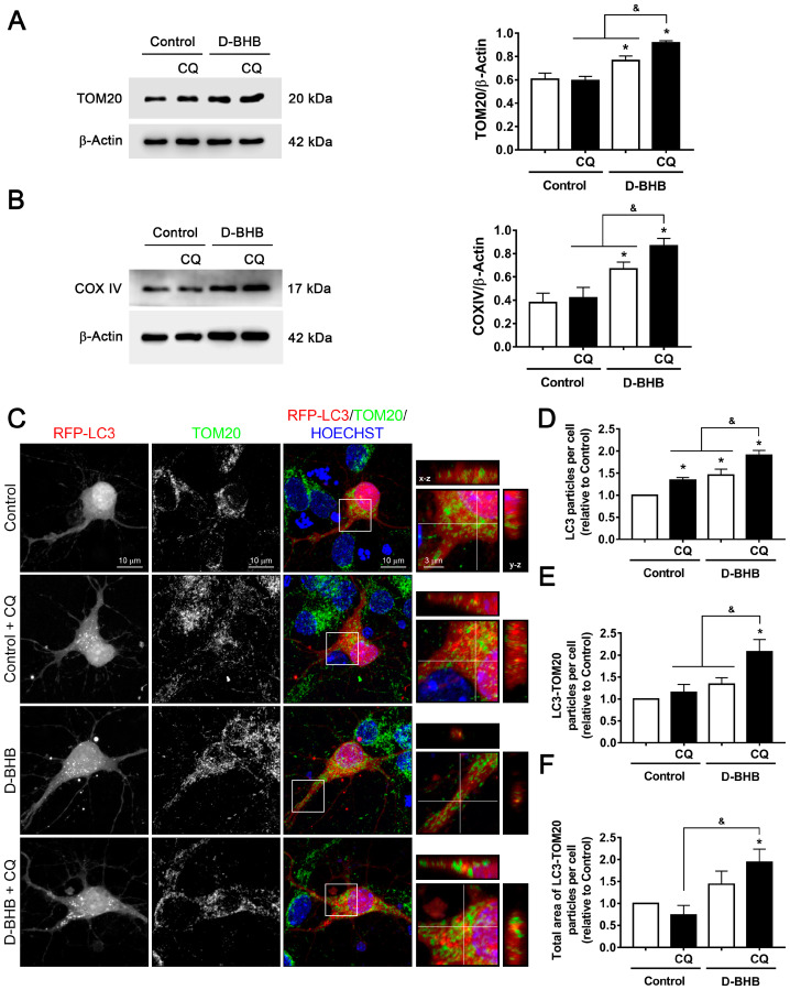
Effect of D-BHB on mitophagy in cortical neurons by colocalization of RFP-LC3 and TOM20. Representative Western blot and quantification of (A) TOM20/β-actin and (B) COXIV/β-actin from cortical neurons treated and non-treated with D-BHB (5 mM) for 24 h, with or without CQ (20 μM) for 4 h. (C) Representative images of neurons transfected with RFP-LC3 (red) and immunofluorescence against TOM20 (green), incubated or not with D-BHB (5 mM) for 24 h, and treated or non-treated with CQ (20 µM) for 4 h. Inserts show LC3-TOM20 positive particles in orthogonal y–z (right) and x–z (top) projections. Hoechst (blue) was used as nuclei marker. (D) Number of RFP-LC3-positive particles per cell. (E) Number of LC3-TOM20-positive particles per cell. (F) Total area of LC3-TOM20-positive particles per cell. Data are expressed as Mean ± SEM from 4 independent experiments. Data were analyzed by two-way ANOVA followed by Fisher’s post-hoc test. * p < 0.05 vs. control; & p < 0.05 vs. D-BHB + CQ.

