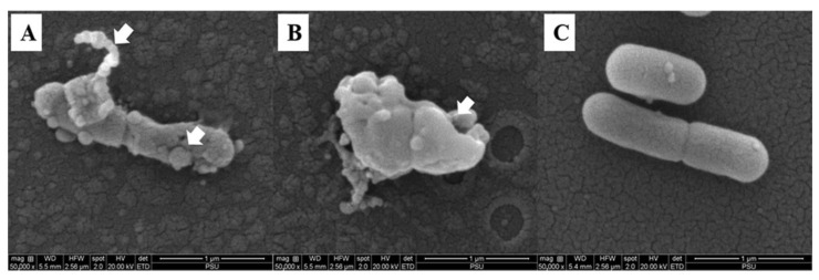
Figure 8.
Scanning electron microscopic (SEM) images of L. monocytogenes treated with 4 × MIC of COS-EGCG conjugate (A,B) and treated with 0.01% acetic acid (negative control) (C). The magnification was 50,000×. Arrow signs indicate the distortion of flagella and rough surfaces (A) and cell surface damage with pores (B).

