Figure 10.
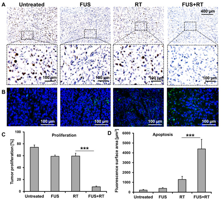
The prostate cancer xenograft tumor showed suppressed proliferation and enhanced apoptosis after FUS + RT compared to single RT. (A) Representative immunohistochemistry staining and (C) quantification of Ki67 demonstrated that the tumor proliferation was inhibited after a combination treatment of FUS and RT. (B) Fluorescence microscopy images of the TUNEL assay and (D) quantified fluorescence surface area show tumor apoptosis after treatment (green: apoptosis; blue: nucleus). N = 4 (*** p ≤ 0.001).

