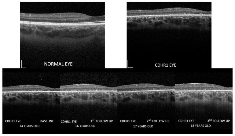
Figure 5.
Images of the inner retina at higher magnification comparing a normal control eye with the CDHR1 eye (top row). Follow-up images recorded at baseline (2019), at the first (2020), the second, and the third follow-up (2022) are shown in the bottom row. Calibration bars indicate 200 micron both vertically and horizontally. Note in the CDHR1 eye the progressively increasing outer retina abnormalities, the inner retina remodeling with loss of normal lamination, specifically the reduced visibility of hyporeflective layers such as ONL and INL, and the progressive increase in the thickness of the inner retina. No differences in ONL/INL thickness between nasal and temporal macula were observed.

