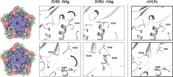
Figure 5: Structural characterisation of rsVLPs.
Depiction of different conformations observed with NAg to HAg shift, and the loss of pocket factor. Cartoon and stick representation of the native conformation (PDB-3VBS), expanded conformation (PDB-3VBU) and rsVLP (major population) focusing upon the region surrounding the canyon and pocket. Residues associated with EC VLP stabilisation in rsVLPs are highlighted: VP3 I325M, VP1 Y116C, VP1 K162I, and the interacting residues VP1 D110, and VP3 D183. Some regions of the capsid have been clipped for clarity.

