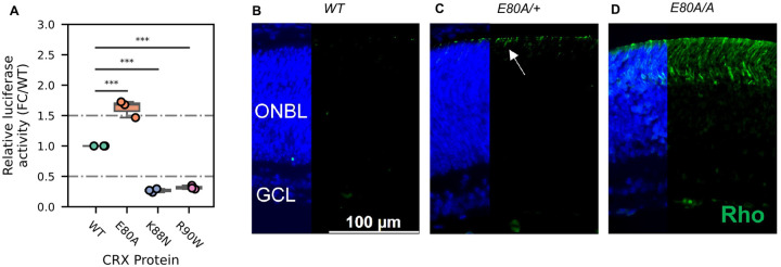
Figure 6. CRX E80A hyperactivity underlies precocious photoreceptor differentiation in CrxE80A retinas.
(A) Boxplot showing luciferase reporter activities of different CRX variants. P-values for one-way ANOVA with Turkey honestly significant difference (HSD) test are indicated. p-value: ****: ≤0.0001, ***: ≤0.001, ns: >0.05. (B-D) Rhodopsin (RHO, green) immunostaining is absent in P3 WT retina but detected in CrxE80A/+ and CrxE80A/A retinas. Nuclei are visualized by DAPI staining (Blue). Arrow indicates the sporadic RHO staining in CrxE80A/+ sample. ONBL: outer neuroblast layer; GCL: ganglion cell layer. Scale bar, 100μm.

