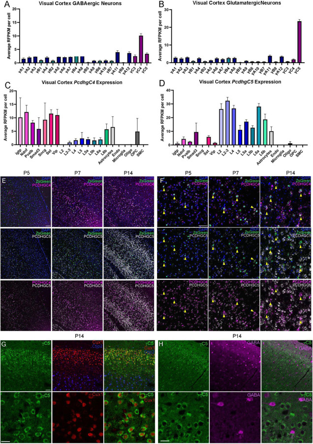
Figure 1. Pcdhγ C-type expression in the mouse cortex during programmed cell death.
A-B. ScRNA-seq from previously generated dataset show expression bias of Pcdhγc4 in GABAergic cIN (A), and Pcdhγc5 in Glutamatergic neurons (B).Error bars represent the error of the mean.
C-D. Expression levels of Pcdhγc4 is higher in inhibitory neuron subtypes (C), while Pcdhγc5 expression is higher in Glutamatergic neuron subtypes (D). Error bars represent the error of the mean. interferon gamma-induced GTPase (Igtp), Neuron-derived neurotrophic factor (Ndnf), Parvalalbumin (Pvalb), Somatostatin (SST), and Vasoactive intestinal peptide-expressing (Vip).
E-F. RNA scope of Nkx2.1;Ai6 cortex during programmed cell death. Low magnification (E) and higher magnification (F) shows expression of Pcdhγc4 and Pcdhγc5 to be minimal at Pcdhγc5, Pcdhγc4 expression is increased by P7 and is expressed in MGE-derived ZsGreen+ cells (yellow arrows). At P14, expression of both Pcdhγc4 and Pcdhγc5 is high, but distinctly not in the same cells, with Pcdhγc4 being highly expressed in ZsGreen+ MGE-derived cINs. Scale bar low-magnification (G) = 100 um. Scale bar high-magnification (F) = 50 um.
G-H. IHC in P14 mouse cortex shows Pcdhγc5 association with excitatory neurons (Ctip2 and Cux1)(G), and not with GABAergic cIN (H). Scale bar top row = 50 um. Scale bar bottom row = 25 um.

